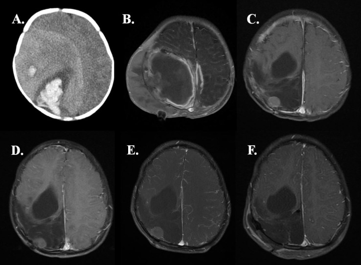
Figure 1.

(A) Axial CT head at 3 months old from initial presentation showed right hyperdense lesion with acute haemorrhage, vasogenic oedema and midline shift from initial presentation. (B) T1-post contrast MRI brain showed gross total resection 9 days postoperative from first resection. (C) T1-post contrast MRI brain at 10 months old a month postoperative from endoscopic cyst fenestration and second resection showed a 1.5 cm rim-enhancing nodule in right parietal region. (D) Surveillance T1-post contrast serial MRI showed interval growth of new lesion at 2 years old and (E) 4 years old with final measurement 1.7 cm. (F) Postoperative T1 MRI after third resection shows gross total resection of the nodule.
