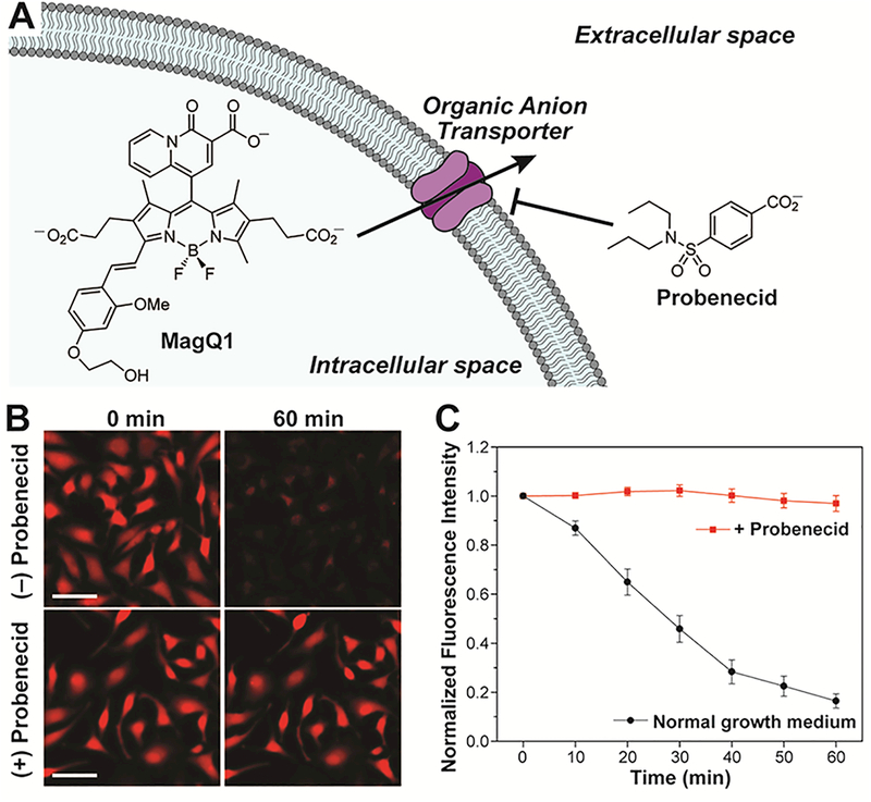
Figure 5.

(A) Possible mechanism of cellular extrusion of anionic dyes by organic anion transporters. (B) Representative widefield microscopy images of live HeLa cells stained with MagQ1, imaged in normal growth medium (top) or medium treated with probenecid, an OAT inhibitor (bottom). Scale bar = 50 μm. (C) Normalized average fluorescence per cell in samples stained with MagQ1, imaged in normal growth medium (black squares), and medium treated with probenecid (red circles). Error bars represent the SD, N = 40.
