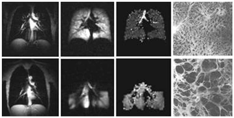
FIG. 2.

Images of normal and emphysematous human lungs. Left to right–proton MRI, 3He ventilation maps, 3He gas ADC maps and histological slices (the latter adapted from Ref. 122); first row–normal lungs, second row–lung with emphysema. ADC in a normal lung is rather homogeneous except for large airways (trachea and its first branches) and is about 0.17 cm2/s. In the emphysema lung 3He gas penetrates only into ventilated regions (lower portion of the lung in this case) and has an ADC about three times bigger (0.55 cm2/s) than the ADC in the normal lung.
