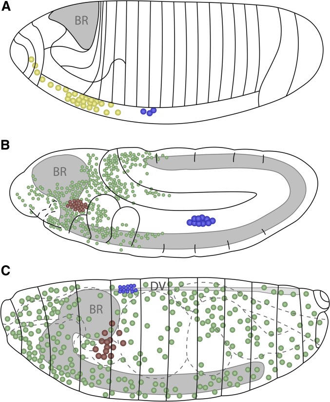
Figure 2.
Embryonic hematopoiesis. (A) Stage 5 embryo. Precursors for embryonic hemocytes (yellow) are specified from the head mesoderm, while lymph gland precursors (blue) arise from the thoracic region of the dorsal mesoderm. BR, gray. (B) Stage 11 embryo. Embryonic prohemocytes migrate and differentiate into plasmatocytes (green) and crystal cells (red). The lymph gland anlage proliferate and are seen in the trunk region. (C) Stage 17 embryo. Plasmatocytes migrate throughout the embryo, while crystal cells accumulate near the proventriculus. During dorsal closure, the lymph gland precursors on either side of the embryo move dorsally and are positioned flanking the DV. Later, these cells will constitute the lymph gland with pairs of distinguishable primary and posterior lobes. Schematics in (A–C) adapted from Volker Hartenstein, see Lebestky et al. (2000). BR, brain; DV, dorsal vessel.

