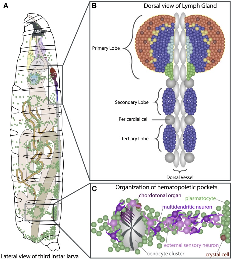
Figure 3.
Larval hematopoiesis. (A) In the third-instar larva, the LG lobes are positioned spanning the DV (gray) posterior to the BR. Plasmatocytes (green) and crystal cells (red) circulate in the hemolymph throughout the larva and are also seen in segmentally distributed sessile pools. (B) The lobes of the LG flank the DV. The primary lobes are the largest and most anterior within the LG, and consist of several distinct cell types and zones (see Figure 4). The posterior lobes (blue) are smaller and remain largely undifferentiated. Pericardial cells (gray spheres) separate the individual lobes of the LG. (C) Detailed view of sessile hematopoietic pockets. The majority of sessile hemocytes, including plasmatocytes and crystal cells, reside along the dorsal side of the larva in stereotypically arranged lateral patches termed hematopoietic pockets. Clusters of oenocytes (gray) also reside within these regions, but are dispensable for the formation of the sessile pools. Activity of external sensory and multidendritic neurons (purple) is necessary for adherence, proliferation, and maintenance of hemocytes within hematopoietic pockets. Schematic in (A) adapted from Volker Hartenstein, in (C) adapted from Gold and Brueckner (2015). BR, brain; DV, dorsal vessel; EA, eye-antennal disc; LG, lymph gland; MH, mouth hooks; PV, proventriculus; SG, salivary gland.

