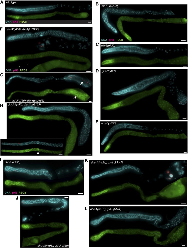
Figure 2.
Genetic evidence that dlc-1 functions with gld-1. Dissected gonads of the indicated genotypes were stained for the M-phase marker phosphohistone H3 (pink) and DNA (blue). Stem and progenitor cells are detected by anti-REC-8 staining (green). (A) Wild-type control cultured at 20°. Single mutant animals including (B) dlc-1(tm3153), (C) gld-3(q730), (D) gld-2(q497), and (E) nos-3(q650) show no ectopic germ cell proliferation. Aberrant proliferation (white arrows) is observed in (H) gld-2(q497); dlc-1(tm3153) and (G) gld-3(q730); dlc-1(tm3153) double mutant animals (inset depicts a gld-2(q497); dlc-1(tm3153) mutant germline with >35 stem and progenitor cell rows), but not in (F) nos-3(q650); dlc-1(tm3153). No ectopic proliferation is detected in (I) dhc-1(or195) single mutant animal, (J) dhc-1(or195); gld-3(q730) double mutant animal, (K) dhc-1(js121) mutant treated with control RNAi, or (L) dhc-1(js121); gld-2(RNAi). Efficacy of gld-2(RNAi) was determined by scoring sterility and embryonic lethality of dhc-1(js121)/hT2; gld-2(RNAi) treated worms. dhc-1(js121)/hT2; gld-2(RNAi) treated worms exhibited 20 ± 6% sterility and for worms with progeny the embryonic lethality was 98 ± 0.9%. Fluorescence micrographs are representative images of data collected from at least two independent experiments and 19–80 worms were scored for each genotype (see Table 1). Bar, 10 μm.

