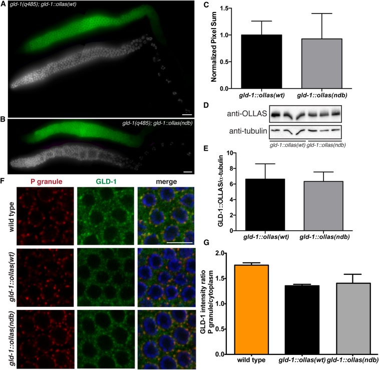
Figure 8.
GLD-1ndb mutation does not affect protein expression and localization in vivo. GLD-1wt::OLLAS and GLD-1ndb::OLLAS protein expression (green) in (A) gld-1(q485); gld-1wt::ollas and (B) gld-1(q485); gld-1ndb::ollas transgenic worm gonads. GLD-1::OLLAS is highly expressed in the meiotic pachytene region of the germline. DNA was stained using DAPI (gray) for reference. Bar, 10 μm. (C) Quantitation of GLD-1::OLLAS immunostaining intensity levels in meiotic pachytene cells using LAS-X software (Leica). No significant difference in expression was observed using Student’s unpaired t-test (P > 0.6; gld-1wt::ollas N = 12, gld-1ndb::ollas N = 13). (D) Western blot analysis of GLD-1wt::OLLAS and GLD-1ndb::OLLAS protein levels in whole lysates of transgenic worms. Tubulin is used as a loading control. (E) Quantitation of GLD-1wt::OLLAS and GLD-1ndb::OLLAS protein level in 50 transgenic whole worm lysate normalized to tubulin. No significant difference in expression was detected by Student’s unpaired t-test (P > 0.7; N = 5 for each genotype). (F) Confocal images of meiotic pachytene region of wild-type, gld-1(q485); gld-1wt::ollas, and gld-1(q485); gld-1ndb::ollas germlines co-immunostained for P granule component PGL-1 (red) and GLD-1::OLLAS (green). Bar, 10 μm. (G) Quantitation of GLD-1, GLD-1wt::OLLAS and GLD-1ndb::OLLAS protein enrichment in P granules from confocal images. No significant difference in enrichment of GLD-1wt::OLLAS and GLD-1ndb::OLLAS was detected by Student’s unpaired t-test (P > 0.6; N = 3 for each genotype).

