Figure 9.
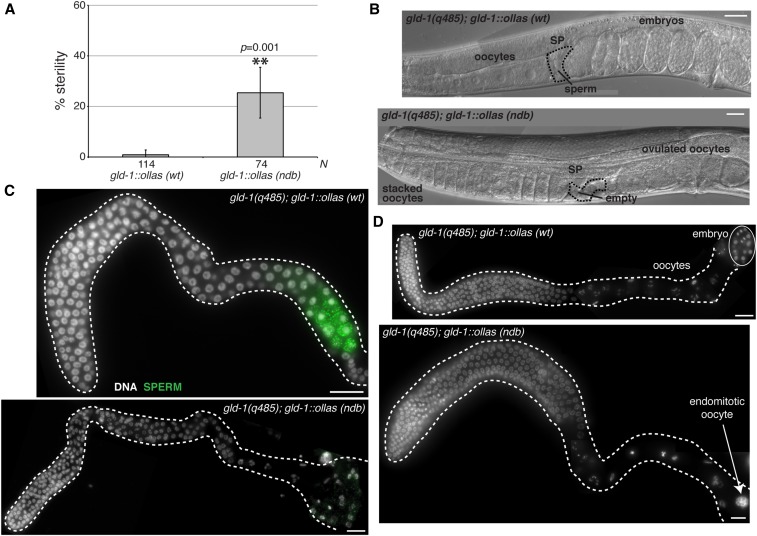
DLC-1/GLD-1 interaction promotes GLD-1 function in vivo. (A) Analysis of sterility phenotype of gld-1(q485); gld-1wt::ollas and gld-1(q485); gld-1ndb::ollas worms at 24°. Sterile worms were identified by the inability to produce viable offspring. L4 larvae from synchronous cultures of either gld-1(q485); gld-1wt::ollas or gld-1(q485); gld-1ndb::ollas worms were isolated and their ability to produce viable offspring assessed. Percent sterility from four independent experiments was calculated and error bars represent SD from the mean. Number of animals scored for each genotype (N) is indicated below the chart. ** indicates statistically significant difference in percent sterility determined by Student’s paired t-test (P = 0.001). (B) Images of gld-1(q485); gld-1wt::ollas and gld-1(q485); gld-1ndb::ollas whole worms acquired using Nomarski DIC microscopy. Morphological landmarks include oocytes, spermatheca (SP; black dashed outline), sperm and embryos. gld-1(q485); gld-1ndb::ollas worms accumulate oocytes, ovulate unfertilized oocytes and spermatheca appears empty. (C) Fluorescence micrograph of gld-1(q485); gld-1wt::ollas or gld-1(q485); gld-1ndb::ollas gonads dissected at L4 stage and immunostained for sperm (green) using anti-MSP antibody. A subset (8%) of gonads from gld-1(q485); gld-1ndb::ollas worms lack sperm and exhibit feminized germline phenotype (Table 2). (D) Dissected gonads from either gld-1(q485); gld-1wt::ollas or sterile gld-1(q485); gld-1ndb::ollas worms stained with DAPI to reveal DNA morphology. Gonads from gld-1(q485); gld-1ndb::ollas mutant worms exhibit endomitotic oocyte phenotype (white arrow) (27%; Table 2). Bar, 10 μm.

