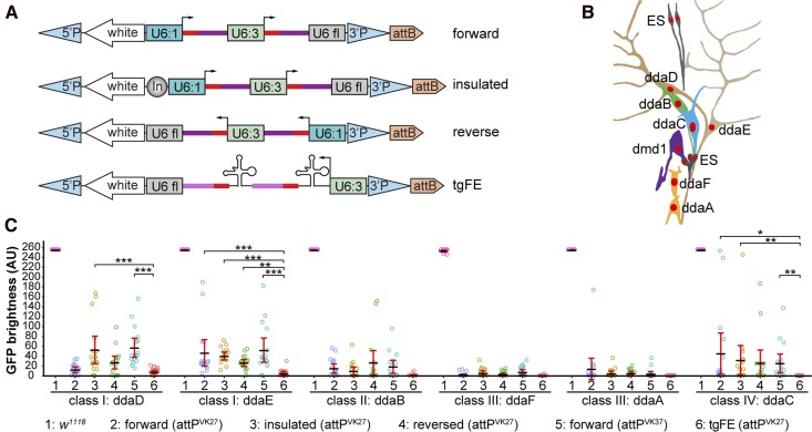
Figure 2.
Optimization of multi-guide RNA (gRNA) design for tissue-specific gene knockout in Drosophila. (A) Four designs of multi-gRNA transgenic vectors. U6:1 and U6:3, U6 promoters; U6 fl, U6 3′ flanking sequence; In, Gypsy insulator. Red bars, gRNA targeting sequence; dark magenta bars, original gRNA scaffold; light magenta bars, E+F gRNA scaffold. (B) Diagram of the dorsal cluster of larval peripheral sensory neurons. (C) Comparison of a control (1) and various gRNA-GFP lines in eliminating GFP signal in each dorsal da neuron using RluA1-Cas9. Da neurons express UAS-CD8-GFP driven by nsyb-Gal4. The integration site for each gRNA line is indicated in parentheses. The GFP signals in most control neurons are saturated under the setting used. Each circle represents an individual neuron (n = 16 for each column). Black bar, mean; red bars, SD. * P ≤ 0.05, ** P ≤ 0.01, ***P ≤ 0.001; one-way ANOVA and Tukey’s honest significant difference test. Only significance levels between #6 and others are indicated. AU, arbitrary units.

