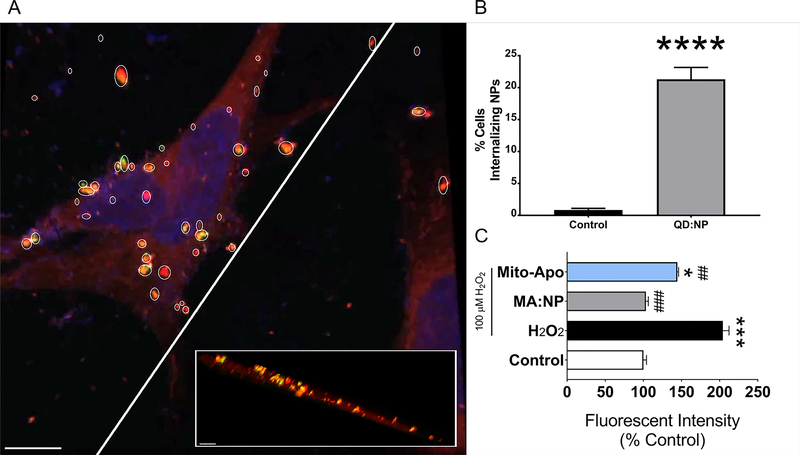
Figure 4.
NP internalization and protection in N27 neurons. (A) Confocal microscopy of 30 μg/mL 1% QD:NPs (yellow)-loaded N27 cells in 2% RPMI for 24 h before staining for mitochondria (red; MitoTracker® red dye) and nucleus (blue; hoechst) and fixing. The cross-sectional perspective is shown by the slice in the main image. Scale bar: 10 μm. Inset scale bar: 5 μm. (B) Flow cytometric analysis of N27 cells. Cells were incubated with 30 μg/mL 1% QD:NPs for 24 h in 2% RPMI. Cells were then collected, washed once with PBS to remove excess NPs and fixed with 4% PFA. Fluorescence was measured using a 633 nm laser, using unloaded cells for gating. **** p<0.0001 with respect to control. (C) Protection of cells treated with 30 μg/mL 0.2% MA:NPs against oxidative stress using a caspase-3 assay. After incubating cells for 18 h with either 30 μg/mL MA:NPs or 5 μg/mL (=10 μM) mAPO, media was replaced with 2% RPMI containing 100 μM H2O2 as well as the same concentration of MA:NPs or free mAPO, and incubated for 6 h. After the 6 h challenge, cells were collected and lysed using a caspase-3 buffer; supernatants from lysate were incubated with caspase-3 substrate for 1 h and fluorescence was quantified (em: 380, ex: 460 nm) using a 96-well plate reader. * p<0.05 with respect to control, *** p<0.001 with respect to control, ## p<0.01 with respect to H2O2, ### p<0.001 with respect to H2O2.

