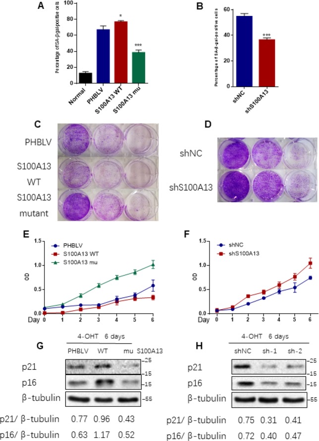
Figure 4.
S100A13 overexpression enhances Ras OIS, while S100A13 silencing attenuates Ras OIS. (A and B) Normal IMR90 cells were infected with lentivirus carrying oncogenic RAS to induce OIS, and at the same time cells were co-infected with (A) PHBLV, S100A13 wild type or mutant type, or (B) control vector (shNC), shRNA against S100A13. After 6 days, cells were stained for SA-β-gal activity. The percentages of cells positive for SA-β-gal were calculated and graphed (n=3). Error bars represent means ± SD from three independent experiments. *P < 0.05, **P < 0.01, ***P < 0.005. (C and D) IMR90 cells were infected as (A) and (B) and cultured for 10–14 days. Then, colony formation assay was performed. (E and F) IMR90 cells were infected as (A) and (B), and cell growth curves were determined by CCK-8/WST-8 assay for the indicated time. Values represent the means ± SD of triplicate points from a representative experiment (n = 3), which was repeated three times with similar results. (G and H) ER:Ras IMR90 cells were infected as (A) and (B), and were given 4-OHT for 6 days. Then, the indicated proteins were detected by Western blot analysis.

