Figure 3.

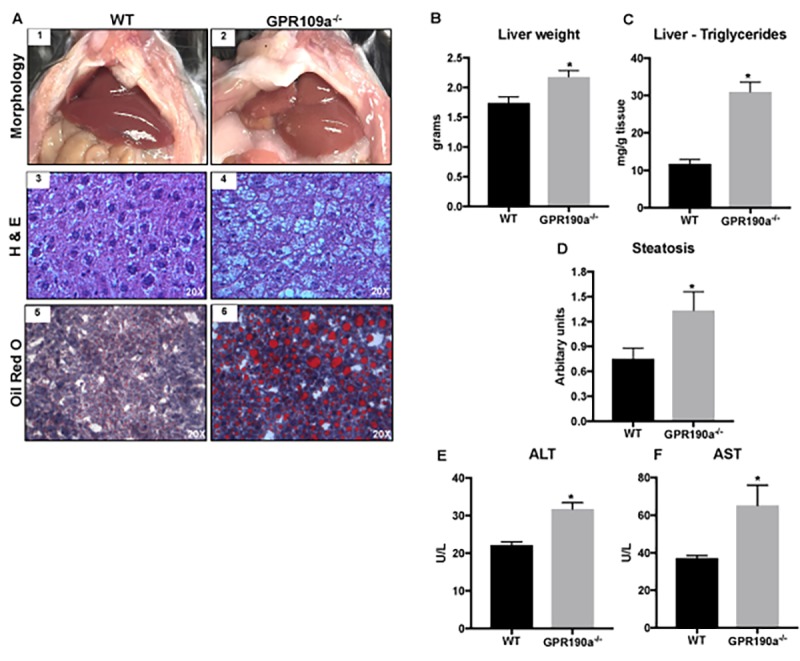
Absence of GPR109A induces age-associated hepatic steatosis in mice. (A) Morphological appearance (1-2), hematoxylin and eosin (3-4) and Oil red O staining of 12 month old WT and Gpr109a-/- mouse liver sections (20X) (5-6). (B) Changes in liver weight and (C) triglyceride content of 12-month-old WT and Gpr109a-/- mice. (D) Semi-quantitative scoring system was used to assess hepatocyte steatosis (0, 1-5% of total area; 1, 5 – 33% of the total area; 2, 33 – 66% of the total area; 3, >66% of the total area; results represented in arbitrary units. (E-F) Changes in the circulating levels of liver injury markers. Data is represented as mean ± S.E.M for (n=6). *p<0.05 vs. WT.
