
Extended Data Figure 6. B cells are dispensable for the altered metabolic phenotypes in integrin β7 deficient mice.
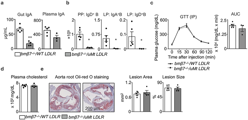
Ldlr−/− mice were lethally irradiated and reconstituted with BM cell mixtures of β7−/− and WT (β7−/−/ WT, 1:1 ratio), or, β7−/− and μMt (β7−/−/μMt, 1:1 ratio). The reconstituted mixed chimeras were fed on high-cholesterol diet (HCD) for 14 weeks. a, IgA levels in the gut flush (n = 5 for β7−/−/ WT vs n = 4 β7−/−/ μMt mice, mean ± s.e.m) and the plasma (n = 5 for β7−/−/ WT vs n = 3 β7−/−/ μMt mice, mean ± s.e.m), **P < 0.01, Mann-Whitney two-tailed test. b, Number of IgD+ B cells in Peyer’s patches and IgA+ B cells and IgD+ B cells in lamina propria as determined by flow cytometry (n = 5 for β7−/−/ WT vs n = 3 β7−/−/ μMt mice, mean ± s.e.m), *P < 0.05, Mann-Whitney two-tailed test. c, Glucose tolerance test in HCD-fed mixed chimeras (n = 5 for β7−/−/ WT vs n = 3 β7−/−/ μMt mice, mean ± s.e.m). d, Plasma cholesterol levels in overnight-fasted mice (n = 5 for β7−/−/ WT vs n = 4 β7−/−/ μMt mice, mean ± s.e.m). e, Representative images and quantification of Oil-red O staining in aorta root sections of bmβ7−/−/ WT Ldlr−/− and bmβ7−/−/ μMt Ldlr−/− mice on HCD for 14 weeks (n = 5 for β7−/−/ WT vs n = 4 β7−/−/ μMt mice, mean ± s.e.m).