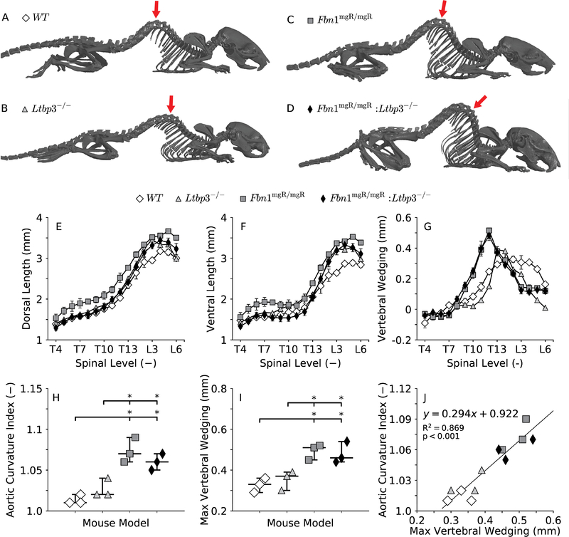
Figure 4.
Representative images of skeletal reconstructions for all four genotypes (A-D). Note that the mgR mutation, even in the absence of LTBP-3, predisposes to excessive thoracic kyphosis, which might be related to the abnormal residual shape of the DTA in Fbn1mgR/mgR and Fbn1mgR/mgR:Ltbp3−/− mice (Figure 3, C-D). The severity of thoracic kyphosis is measured in a position-independent manner (Li et al. 2017) through ventral wedging of the vertebrae (G), calculated as the difference between the dorsal (E) and the ventral (F) length of the vertebral bodies in the thoracic (vertebrae T4-T13) and lumbar (vertebrae L1-L3) spine. The aortic curvature index (H), calculated as the ratio of the contour length to the end-to-end distance between the left subclavian and the sixth pair of intercostal arteries (Figure 3, D), quantifies the extent to which the profile of control and mutant DTAs deviates from a straight line. Maximum spinal wedging (I), that is, the maximum difference between the lengths of the dorsal and ventral aspects of the vertebrae (G), quantifies the maximum degree of spinal deformity in the thoracic (vertebrae T4-T13) and lumbar (vertebrae L1-L3) spine (its anatomical location shown by red arrows in panels A-D). The deviation of the DTA from a straight line (H) and kyphosis of the thoracic spine (I) are more pronounced in both the Fbn1mgR/mgR and Fbn1mgR/mgR:Ltbp3−/− mice than in the control mice. In fact, a strong significant correlation exists between these two parameters (J), suggesting that the configuration of the growing spine influences the natural shape of the aorta. Statistical significance is set to *p < 0.05.

