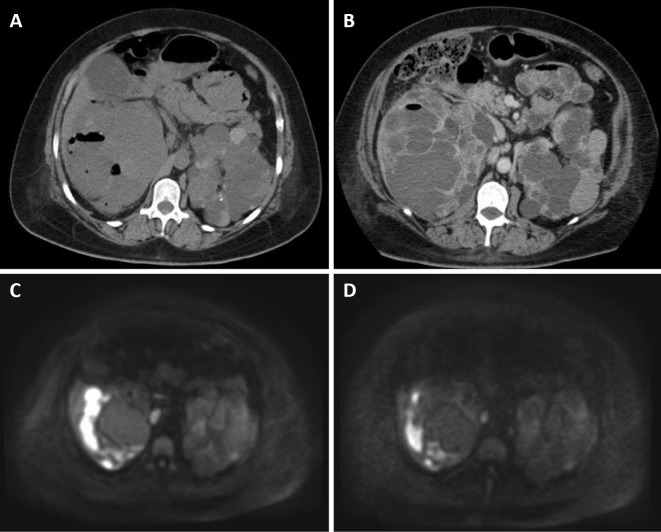
Figure 2.
Abdominal computed tomography (CT) and magnetic resonance imaging (MRI). A: Abdominal CT on the 3rd hospital day, showing gas within the cyst in the right kidney. The cysts on the upper pole of the right kidney are poorly defined and gaseous with opacity in the surrounding fat tissue and peritoneal thickening, diagnosed as cystic infection. B: Abdominal contrast-enhanced CT on the 9th hospital day, still showing gas within the cyst in the right kidney. The cysts became less gaseous after the administration of antibiotics, but infected cysts remained. C: Abdominal MRI (diffusion-weighted image) on the 18th hospital day, showing high intensities in the right kidney. D: Abdominal MRI (diffusion-weighted image) on the 35th hospital day, still showing high intensities in the right kidney.

