Figure 1.
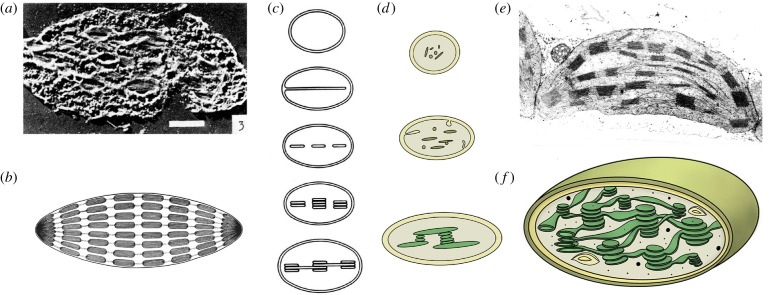
Chloroplast structure and development in the past and today. Already at the beginning of the 1950s, the first electron microscopic pictures of chloroplasts of land plants were taken. (a) The first cross-section through a tulip chloroplast [14]. (b) The inside of chloroplasts was considered to consist of stroma and connected grana thylakoids that were believed to be arranged like rolls of coins [15]. (c) Differentiation towards mature chloroplasts was suggested to happen from a progenitor via invaginations of the inner envelope. Invaginations would then split into disc-shaped vesicles that stacked together to be eventually interconnected [16]. (d) The modern view on chloroplast differentiation is very similar to that of the past. The progenitors are proplastids that contain only few internal membranes and vesicles that finally assemble the thylakoid membrane network in the presence of light. (e) Modern electron micrographs provide insights into the actual arrangement of the grana stacks. The complex internal organization of a chloroplast is depicted in (f) with thylakoids forming a highly interconnected fretwork.

