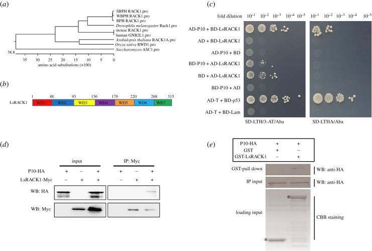
Figure 1.
Interaction between RBSDV P10 and LsRACK1. (a) Phylogenetic analysis using RACK1 amino acid (aa) sequences from different sources. (b) Schematic of RACK1. WD40 domains are short structural motifs with about 40 aa and the numbers above the diagram represent the aa positions. (c) Results of yeast two-hybrid assays. RBSDV P10 and LsRACK1 were cloned individually into pGADT7 (AD) and pGBKT7 (BD) vectors. Serial dilutions of yeast cells co-transfected with two various combined vectors were plated on the SD-LTH/3-AT/Aba or SD-LTHA/Aba medium. Cells co-transfected with AD-T and BD-p53 and with AD-T and BD-Lam were, respectively, used as a positive and a negative control. Positive interactions are indicated by the growth of the cells. (d) Co-IP. LsRACK1-Myc and P10-HA were separately or co-expressed in Sf9 cells by transfection. LsRACK1-Myc expressed alone in cells served as a negative control. At 48 h post transfection, Sf9 cell lysates were immunoprecipitated with anti-Myc beads followed by Western blot (WB) using an anti-HA (upper panel) or an anti-Myc antibody (lower panel). (e) GST pull-down assay. GST or GST-LsRACK1 was incubated with 6X His-P10-HA and then mixed with glutathione-sepharose beads. The beads were washed and analysed by Western blot using an anti-HA antibody (upper panel). The middle panel shows the Western blot results of inputs of HA-tagged P10 proteins from pull-down assays. Equal volume of glutathione-sepharose beads carrying GST-LsRACK1 or GST were analysed by SDS-PAGE and stained with Coomassie blue. The position of GST-LsRACK1 or GST is indicated with asterisks.

