Figure 2.
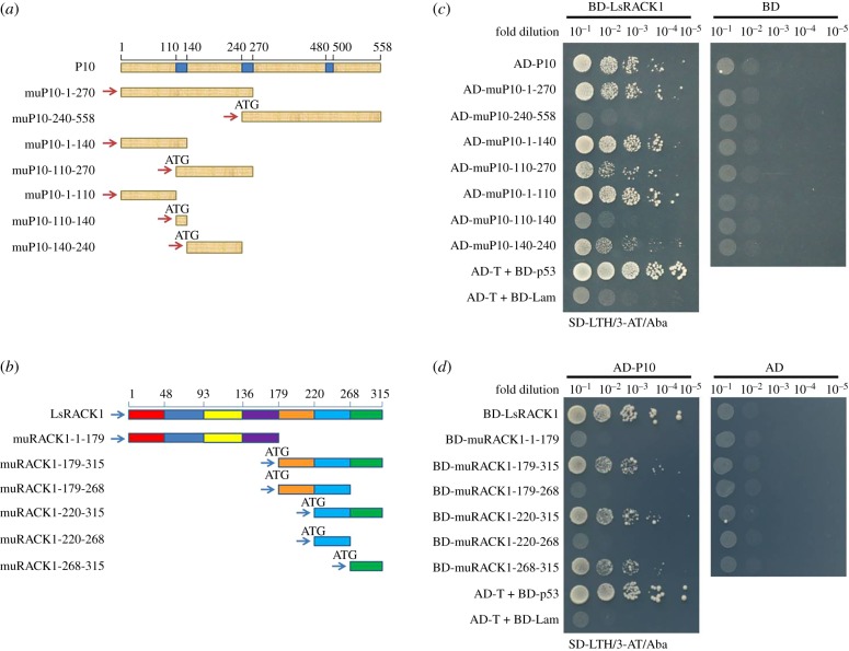
Determination of domains needed for RBSDV P10 and LsRACK1 interaction. (a) Schematic of RBSDV P10 and its deletion mutants. The three blue boxes inside the diagram represent transmembrane domains in the RBSDV P10. The numbers above the diagram represent the positions of amino acids. The seven short fragments below the P10 diagram represent the seven deletion mutants and their respective positions. (b) Schematic of LsRACK1. Different colours shown in the diagram represent seven WD40 domains, respectively. The fragments below the diagram represent the LsRACK1 deletion mutants. (c) Yeast two-hybrid assay results showing the positive or negative interactions between AD-P10 and its deletion mutants with BD-LsRACK1. Co-expression of AD-P10 or its deletion mutants with the BD vector were used as negative controls. (d) Yeast two-hybrid assay results showing the positive or negative interactions between BD-LsRACK1 and its deletion mutants with AD-P10. The cell co-expressing AD-T and BD-p53 was used as a positive control, and the cell co-expressing AD-T and BD-Lam was used as a negative control.

