Figure 1.
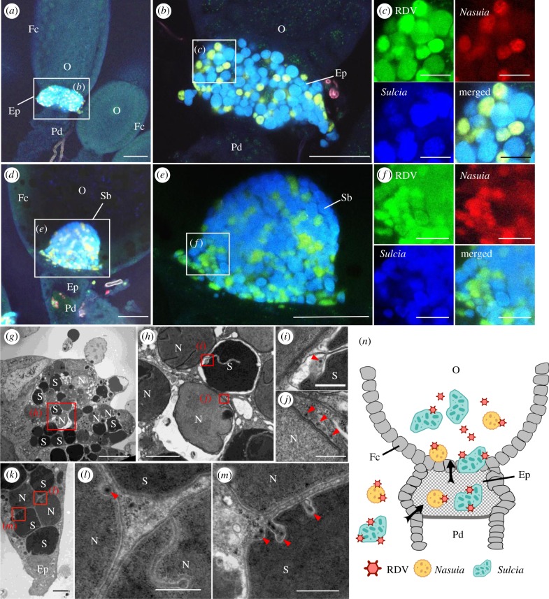
RDV virions moved with the obligate bacterial symbionts Sulcia and Nasuia into the oocyte of female N. cincticeps. (a–c) Confocal micrographs show the colocalization of RDV with Sulcia or Nasuia in the epithelial plug of female insects at 6 days post-emergence. Panel b is an enlargement of boxed area in a. Panels in c are enlargements of boxed area in b. (d–f) Colocalization of RDV and Sulcia or Nasuia in the symbiont ball in the oocyte of female insects at 8 days post-emergence. Panel e is an enlargement of boxed area in d. Panels in f are enlargements of boxed area in e. Scale bars in a, b, d and e: 100 µm; c and f: 25 µm. (g–m) Transmission electron micrographs showing RDV particles distributed along the envelopes of Nasuia or Sulcia at the same follicular epithelial cells in the epithelial plug. Panel h is an enlargement of boxed area in g. Panels i and j are enlargements of boxed areas in h. Panels l and m are enlargements of boxed areas in k. Scale bars in g: 10 µm; h and k: 2 µm; i, j, l and m: 500 nm. (n) Model for the exploitation of bacterial symbionts Sulcia and Nasuia by RDV to enter the oocyte. RDV virions directly attach to the envelopes of Nasuia or Sulcia and then enter the oocyte from the epithelial plug along with Nasuia or Sulcia. Ep, epithelial plug; Fc, follicular cell; N, Nasuia, O, oocyte; Pd, pedicel; S, Sulcia; Sb, symbiont ball. Black arrows mark the entry route of virus-associated bacterial symbionts. Red arrows indicate RDV virions. All electron micrographs and immunofluorescence figures are representative of at least three replications.

