Figure 3.
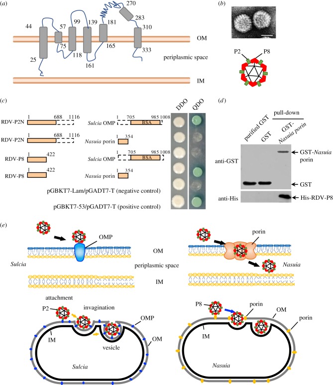
RDV virion entry into Nasuia periplasmic space was mediated by specific interaction between RDV P8 and Nasuia porin. (a) Secondary structure prediction showed that Nasuia porin consisted of 18 β-strands and six transmembrane regions. (b) RDV virion consists of one minor outer capsid protein P2 and one major outer protein P8. Scale bar; 50 nm. (c) Yeast two-hybrid assay to detect interactions between P2N (15 nm domain, 1–688 aa) or RDV P8 and Sulcia OMP (BSA domain, 706–985 aa) or Nasuia porin. (d) GST pull-down assay to detect interactions between RDV P8 and Nasuia porin. Nasuia porin-GST acted as a bait protein with GST as a control. Pull-down samples were probed with GST antibody in a western blot. (e) Proposed model for the different binding strategies for RDV virions with the envelopes of Sulcia and Nasuia. IM, inner membranes; OM, outer membranes; OMP, outer membrane protein; PS, periplasmic space.

