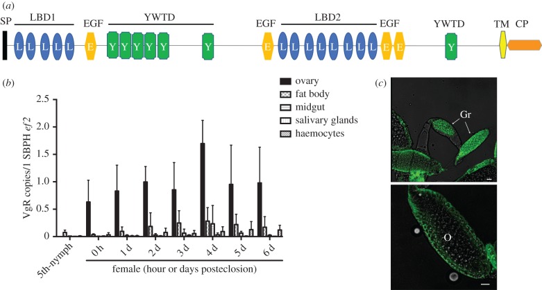
Figure 1.
The Laodelphax striatellus VgR gene and its expression profile. (a) Schematic composition of L. striatellus VgR, as illustrated by the SMART algorithm. SP, signal peptide sequence; LBD, ligand-binding domain; EGF, epidermal growth factor-like domain; YWTD, YWTD amino acid repeats; TM, transmembrane domain; CP, cytoplasmic region. (b) RT-qPCR to quantify VgR mRNA levels in L. striatellus tissues at different developmental stages. Ef2, L. striatellus elongation factor 2 gene. Means and s.d. were calculated from three independent experiments, with five mRNA samples per experiment. (c) Immunofluorescence assay to localize VgR protein in L. striatellus ovaries. VgR was probed with a mouse anti-VgR polyclonal antibody and stained with Alexa Fluor 488 (shown in green). Samples were examined using a Leica TCS SP8 confocal microscope. Images are representatives of three independent experiments each with five insects analysed. Gr, germarium; O, oocyte. Scale bar, 20 µm. (Online version in colour.)

