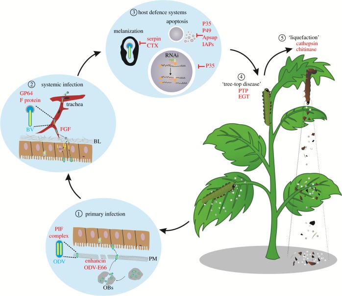
Figure 1.
Model of how baculoviruses overcome different host barriers to establish successful infections. After being ingested by susceptible larvae, OBs are dissolved under the alkaline conditions of the insect midgut to release ODVs. Two enzymes (enhancin and ODV-E66) can degrade the PM to allow the access of ODV to the midgut epithelia. Primary infection is then initiated by a group of ODV-specific envelope proteins (PIFs) (①). To egress from the BL barrier at the basal side of the midgut epithelial cells, baculoviruses use vFGF for the chemotaxis of tracheoblasts to facilitate BV passage, and they use BV-specific envelope proteins (GP64 or F proteins) to spread systemic infection (②). Baculoviruses exploit different strategies to suppress host defence systems, including melanization, apoptosis and RNAi for efficient virus replication (③). Baculoviruses can also regulate host physiology and behaviour, such as inducing a ‘tree-top disease’ via PTP and EGT (④) and ‘liquefaction of infected larval bodies' by chitinase and cathepsin (⑤) for optimal virus dispersal. BL: basal laminae; BV: budded virus; ODV: occlusion-derived virus; OB: occlusion body; PM: petritrophic membrane. Viral proteins are in red.

