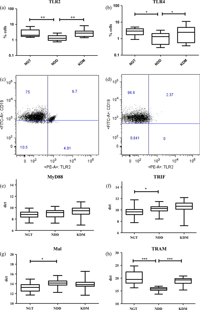
Fig. 1.
Newly diagnosed DM is characterized by decreased surface expression of TLR2 and 4 and deranged expression of TLR adaptors in peripheral blood leukocytes (PBL) as determined by flowcytometry and qRT-PCR respectively. Box and whisker plots showing the levels of expression of TLR2 (a) and TLR4 (b) in NGT/control (n = 13), NDD (n = 14) and KDM (n = 15) subjects. Representative dot plot showing the expression of TLR2+ B cells in NGT (c) and NDD (d) subjects. Box and whisker plots showing the levels of expression of MyD88 (c), TRIF (d), Mal (e) and TRAM (f) in NGT/control, NDD and KDM subjects. Statistical significance was determined by non-parametric Mann–Whitney U test and p < 0.05 was considered significant. NGT – normal glucose tolerance, NDD – newly diagnosed diabetic, KDM – known diabetic subjects.

