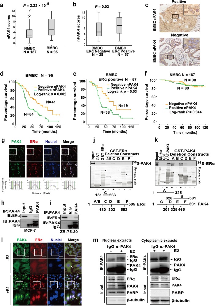
Figure 1.
Nucleus PAK4 is a malignant effector in ERα+ breast cancer with bone metastasis. a Box plot of nPAK4 in the NMBC or BMBC patients. The subjects with breast cancer were divided into two groups based on bone metastasis. b Box plot of nPAK4 in BMBC samples from 95 subjects. The subjects were divided into two groups based on ERα expression scores in the tumors, representing negative and positive for ERα expression. The data were analyzed using the Mann–Whitney U test. The horizontal lines represent the median; the bottom and top of the boxes represent the 25th and 75th percentiles, respectively, and the vertical bars represent the range of the data. c Two representative images showing positive (upper picture) or negative (lower picture) nPAK4 localization in the BMBC samples. Scale bars, 50 µm. d, e Ninety-five cases of BMBC and 57 cases of ERα + BMBC were divided into two groups using the nPAK4 localization signal. The relationship between nPAK4 protein expression and bone metastasis-free survival (BMFS) was analyzed according to the Kaplan–Meier method. P values were obtained using the log-rank test. f PAK4 expression in the nucleus of breast cancer cells was not significantly associated with non-bone relapses (brain, liver, or lung). Kaplan–Meier survival analysis of 187 patients with breast cancer separated into two groups based on the median value of the nPAK4 localization signal. The positive group is shown in green (n = 89), and the negative group is shown in orange (n = 98). P values were calculated using the log-rank test. g Representative images of ERα+ breast cancer tissue (green, PAK4; red, ERα; and blue, nuclei). Scale bar, 20 μm. The 2nd lines are the 2.5-folds enlarged pictures of the 1st lines, respectively. The image-pro plus 6.0 software convert immunofluorescence staining into peaks/curves at a 3rd line across the image. MCF-7 h and ZR-75-30 i cell lysates were immunoprecipitated with PAK4 antibodies or IgG. Then, endogenous ERα and PAK4 were detected using immunoblot assays. j, k For the GST pull-down assay, GST, GST-ERα, GST-PAK4 plus GST-ERα deletions or GST-PAK4 deletions were incubated with the indicated proteins, transcripted, and then translated in vitro. Bound proteins were detected with auto-radiography. A schematic representation of the ERα and PAK4 deletion constructs is shown. l Representative PAK4 and ERα immunostaining in MCF-7 cells treated with or without E2 (10−9 M). PAK4 (green); ERα (red); and nuclei were stained with DAPI (blue). Merged images are shown as indicated. Original magnification: × 40. Scale bar: 37.5 μm. m Co-IP of PAK4 and ERα from the nuclear and cytoplasmic fractions obtained from human MCF-7 cells treated with E2 (10−9 M) for 45 min. β-tubulin and PARP were used as controls for the cytoplasmic and nuclear compartments, respectively

