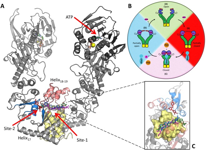
Figure 1.
Illustration of Hsp90α in the open conformation. (A) The location of the different binding site residues are shaded: Site-1 helix18-19 (red), helix21-22 four-helix bundle (yellow) and Site-2 sub-pocket (blue). The NTD location of ATP and magnesium ions (spheres) are also shown. (B) Hsp90’s nucleotide driven conformational cycle (Adopted from Penkler et al.37). (C) Inset – zoomed in view of docked compounds SANC309 red, SANC491 green, SANC518 blue, and Novobiocin magenta.

