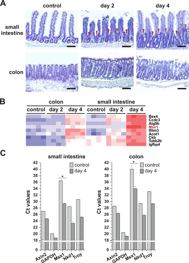
Figure 1.
Msx1 expression is upregulated upon Apc gene inactivation in the mouse intestine. (A) Crypt hyperplasia arising in ApccKO/cKO Villin-CreERT2 small intestine 2 and 4 days after tamoxifen administration. Hematoxylin-stained (blue nuclei) paraffin sections of the small intestine (jejunum) and colon at the indicated time points upon tamoxifen administration are shown. Control tissues were obtained from mice of the same genetic background prior to tamoxifen treatment. Red arrowheads indicate hyperproliferative crypt compartments. Scale bar: 0.15 mm. (B) Expression profiling of ApccKO/cKO Villin-CreERT2 small intestinal and colonic crypt cells 2 and 4 days after tamoxifen administration. Control RNA was isolated from crypt cells with intact Apc. A part of the heatmap is presented showing robust upregulation of the Msx1 gene upon Apc inactivation in both tissues. For a complete list of differentially expressed genes, see supplementary Table S1 (small intestine) and Supplementary Table S2 (colon). (C) Quantitative RT-PCR analysis confirms a significant increase in the Msx1 expression levels upon Apc loss. Total RNA was isolated from ApccKO/cKO Villin-CreERT2 small intestinal and colonic crypts 4 days after tamoxifen administration. Control RNA samples were obtained from the animals treated with the solvent only. The diagrams show threshold cycle (Ct) values normalized to β-actin gene expression (the β-actin gene Ct value in this and other diagrams was arbitrarily set to 17). Axin2, Nkd1, and Troy represent the Wnt/β-catenin-responsive genes. GAPDH was – next to β-actin – used as an additional housekeeping gene. RNA samples obtained from four tamoxifen-treated and four control animals were analyzed; qRT-PCR reactions were run in technical triplicates. The diagrams show representative results obtained from one animal; error bars indicate standard deviations (SDs); *p < 0.05; **p < 0.01.

