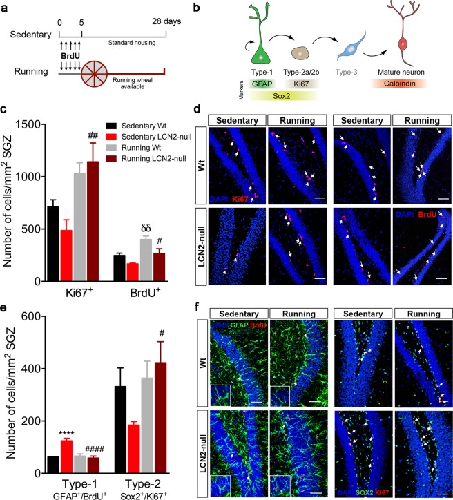
Figure 1.
Voluntary running promotes hippocampal cell proliferation and survival, potentiating the transition of quiescent to proliferating NSCs in LCN2-null mice. (a) Schematic diagram of the experimental paradigm of voluntary running and of the BrdU injection protocol used. For 28 days, Wt and LCN2-null mice were assigned as running, i.e. animals housed to have free access to a running wheel, and as sedentary, housed under standard housing conditions with no running wheel available. (b) Representative illustration of the hippocampal neurogenic process, including cellular types and the specific markers used. (c) Running increased cell proliferation (Ki67+ cells) in LCN2-null mice, and cell survival (BrdU+ cells) in both Wt and LCN2-null mice (n = 4–6 per group). (d) Representative confocal images of Ki67 and BrdU immunostaining (indicated by white arrows) in the SGZ of the DG of Wt and LCN2-null sedentary and running mice. (e) Quantitative analysis of radial quiescent type-1 (GFAP+/BrdU+) and amplifying type-2 stem cells (Sox2+/Ki67+) after 28 days of running revealed a significant decrease in type-1 population in LCN2-null mice, and a consequent increase in type-2 cells (n = 5 mice per group). (f) Representative confocal images of GFAP+/BrdU+ and Sox2+/Ki67+ immunostaining in the SGZ of sedentary and running mice of both genotypes (indicated by white arrows). Scale bars, 50 μm. Data from the sedentary animals is the same as described in Ferreira et al.9, and are presented as mean ± SEM and were analyzed by two-way ANOVA with Bonferroni’s multiple comparison test. *Denotes differences between sedentary Wt and LCN2-null mice; δbetween sedentary and running Wt; #between sedentary and running LCN2-null mice. #p ≤ 0.05, δδ,##p ≤ 0.01, ****,####p ≤ 0.0001.

