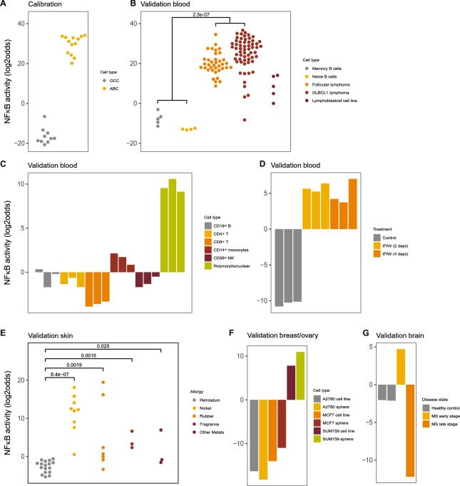
Figure 4.
NFκB pathway model. (A) Calibration of the NFκB pathway model on dataset GSE12195 containing samples from NFκB-active activated B cell-like (ABC) subtype of diffuse large B-cell lymphoma (DLBCL1) and NFκB-inactive germinal centre centroblast (GCC) samples. (B–G) Validation on GSE12195 sample data that were not used for calibration and on independent GEO datasets, various cell/tissue types. (B) GSE12195, healthy B-cells, follicular lymphoma, DLBCL1 with unavailable subtype information, lymphoma cell line. (C) GSE72642, peripheral blood cell types, FACS sorted, of healthy individuals. (D) GSE58096, THP-1 monocytic cells stimulated with IFNγ or vehicle for indicated time periods. (E) GSE60028, patch testing with common antigens (as indicated) and petrolatum (control) was performed on skin of patients with allergic contact dermatitis. (F) GSE43657, breast cancer cell lines, among which the SUM159 inflammatory breast cancer cell line, cultured in 2D and 3D (spheroids) setting. (G) GSE38010, brain tissue samples from early stage (inflammatory) and late stage (inflammation subsided) multiple sclerosis lesions and healthy controls. The activity score is calculated as log2odds. Two-sided Wilcoxon signed-rank statistical tests were performed; p-values are indicated in the figures.

