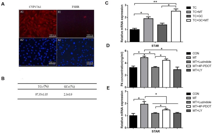
Figure 7.
Effects of melatonin on the relative abundance of STAR mRNA and progesterone production in TCs. (A) Immunohistochemical analysis showed that the CYP17A1 (A1), not the FSHR (B1), expressed in the TCs and DAPI (A2, B2) indicated the nucleus (100×, scale bar = 100 μm). (B) Immunohistochemical analysis indicated that TCs accounted for 97.35 ± 1.05% of all cells. (C) TCs were untreated or treated with 10 ng/mL melatonin and cultured alone or cocultured with GCs for 48 h. (D, E) TCs were treated with 10 ng/mL melatonin alone or in combination with inhibitors, 25 μM LY294002, 10 μM luzindole, or 10 μM 4P-PDOT for 48 h. Total RNA was extracted from TCs. The mRNA expression of STAR was measured by RT-qPCR. GAPDH expression was used as a standard. Progesterone concentration was measured by ELISA. The results are the mean ± SEM of three independent experiments. *P < 0.05, **P < 0.01. LY: LY294002.

