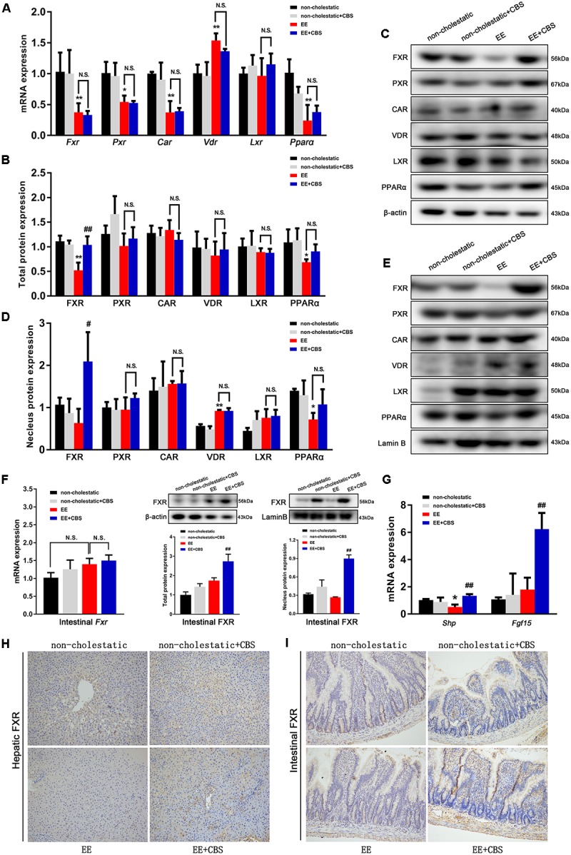
FIGURE 6.

CBS activates the protein expression and nuclear translocation of FXR in liver and intestine. (A) mRNA expression of hepatic nuclear receptors Fxr, Pxr, Car, Vdr, Lxr, and Pparα was determined by real-time PCR and normalized to β-actin. (B,C) Total protein levels and (D,E) nuclear protein levels of hepatic FXR, PXR, CAR, VDR, LXR, and PPARα were determined by Western blot analysis and normalized to β-actin and Lamin B. Representative immunoblot images are shown. (F) mRNA, total protein and nuclear protein levels of intestinal FXR were determined using real-time PCR and Western blot analysis and normalized to β-actin and Lamin B. (G) mRNA levels of Shp in liver and Fgf15 in intestine were evaluated by real-time PCR and normalized to β-actin. Representative images of immunohistochemical staining of (H) hepatic FXR and (I) intestinal FXR. Data are presented as the mean ± SD (n = 6). Significant differences compared with the non-cholestatic group, ∗p < 0.05; ∗∗p < 0.01; compared with the 17α-ethinylestradiol (EE) group, #p < 0.05; ##p < 0.01. N.S., no significance.
