
Fig. 5.
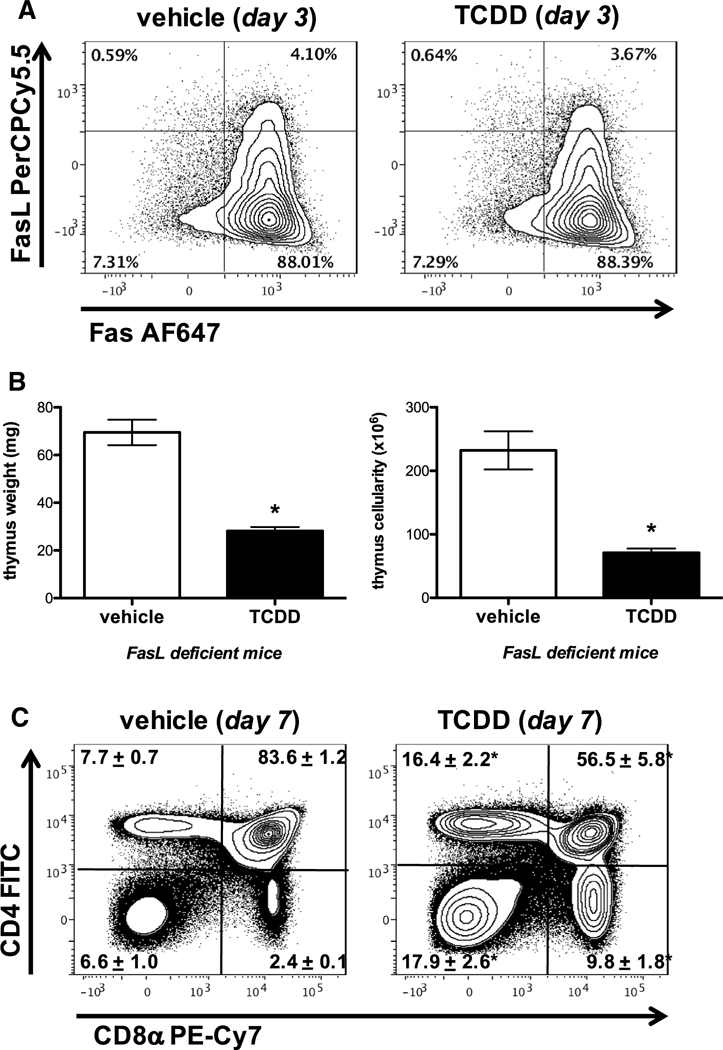
TCDD-mediated thymic atrophy is not dependent on Fas–FasL interactions. Naïve wild-type mice (C57Bl/6) were gavaged with vehicle (anisole/ peanut oil) or TCDD (10 μg/ kg). Representative contour plots gating on singlet thymocytes revealed that 3 days after administration of vehicle or 10 μg/kg TCDD to C57Bl/6 mice, there were no observable effects on the frequency of Fas and FasL expression on CD45+ Thymocytes relative to vehicle control (a). FasL-deficient (gld/gld) mice were exposed to 10 μg/kg TCDD and their thymic weight and cellularity measured on day 7 (b). Similarly, representative dot plots (gating on live thymocytes) revealed a significant decline in the frequency of DP thymocytes, as well as a relative enrichment in the percent of DN and CD4+ CD8− and CD4− CD8+ SP thymocytes in 10 μg/kg TCDD-treated FasL-deficient (gld/gld) mice compared to vehicle control (c). n = 5 mice per treatment group, mean ± SEM; t test, *p < 0.05 vs. vehicle