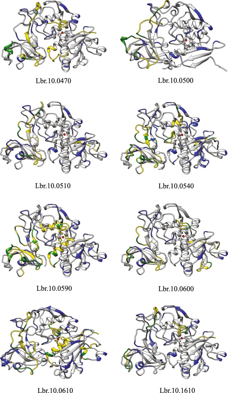
Fig. 7.

Comparative protein modelling of selected Leishmania braziliensis GP63 paralogs from chromosome 10. Modelled structures of eight representative GP63 paralogs from the L. braziliensis chromosome 10. The segments labelled in blue indicate the variable motifs in each protein, while the segments yellow represent the predicted linear B-cell epitopes and the green markings show variable motifs coinciding with the predicted epitopes
