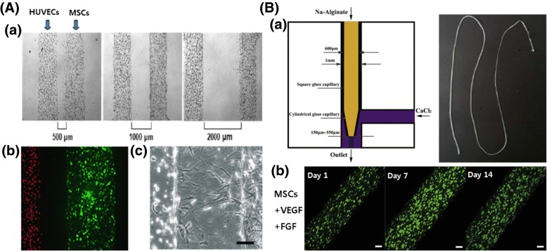
Fig. 6.
Blood vessel regeneration by MSC encapsulated 3D construction. A (a) The 3D co-culture system of mesenchymal and endothelial cells in the micropatterned hydrogel. HUVEC- and MSC-loaded micropatterned fibrin channels with the distances between channels of 500, 1000, and 2000 μm. (b) Encapsulated HUVECs (left channel: red) and MSCs (right channel: green). (c) MSCs sprouted to HUVEC with distance-dependent response (scale bar,100 mm). Reproduced with permission from Reference [36]. Copyright 2009 John Wiley and Sons. B (a) Schematic diagram of microfiber generation and principle of gelation and actual shape. (b) MSCs with VEGF and FGF were effective for angiogenesis in microfiber over 14 days. Scale bar, 200 μm. Reproduced with permission from Reference [65]. Copyright 2017 IOP Publishing

