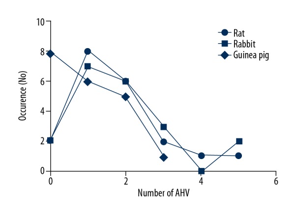
Figure 4.

Comparison of incidence of accessory hepatic veins between laboratory rats, guinea pigs, and rabbits. The occurrence is identified with the number of cases within 1 liver. There was a dominant presence of 1 or 2 accessory hepatic veins in all laboratory animals.
