
Figure 1.
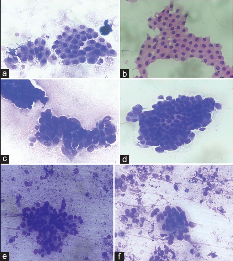
(a and b) Fine-needle aspiration cytology of the gallbladder with diagnostic Category 1 of benign lesions revealing uniform epithelial sheets. (May Grunwald Giemsa, ×40; H and E, ×40). (c and d) fine-needle aspiration cytology of the gallbladder with diagnostic Category 3 of atypical cells showing nuclear overlapping and mild pleomorphism. (May Grunwald Giemsa, ×40; H and E, ×40). (e and f) fine-needle aspiration cytology of the gallbladder with diagnostic Category 4 of atypical cells suspicious of malignancy with few cells showing moderate pleomorphism in necrotic background. (May Grunwald Giemsa, ×40; H and E, ×40)