
Figure 9:
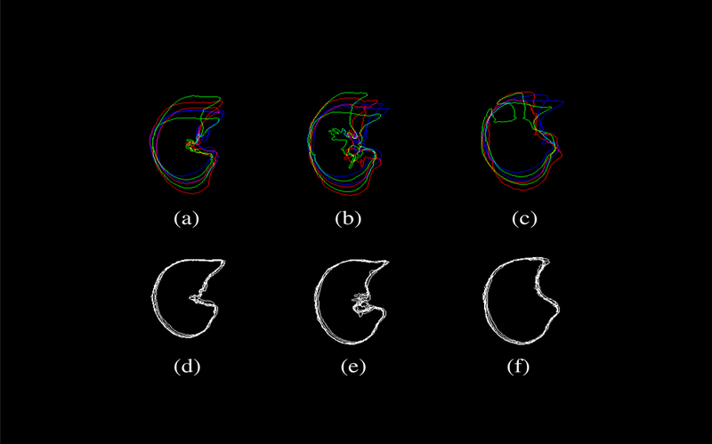
The shape variability among a set of (N=6) subjects involved in this study is demonstrated by overlapping contours from axial CT images. The top row shows ithe shape variation among the subjects, while the bottom row corresponds to the transformed CT datasets after transforming individual subject with respect to a common template subject (subject 9). Only six subjects were displayed here to avoid overcrowding the figure. The columns represent three axial image slices randomly chosen near the apex (top), the body (center), and the diaphragm (bottom). Significant reduction in registration errors can be seen with reduced variability.