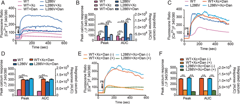
Fig. 2.
Propofol increased cytosolic Ca2+ concentrations ([Ca2+ ]c) more in L286V than WT cells via activation of InsP3 (InsP3R) or ryanodine (RYR) receptors. A) Average [Ca2+]c response to 200 μM propofol (arrow) in WT or L286V cells in the presence or absence of antagonists for InsP3Rs (Xestospongin C, Xc 1 μM) or RYRs (dantrolene or Dan, 30 μM). B) Propofol increased peak [Ca2+]c and integrated Ca2+ calcium response represented by area under the curve (AUC) significantly more in L286V than in WT cells, which were significantly inhibited by Xc or dantrolene alone. C) Average [Ca2+]c response to 200 μM propofol (arrow) in WT or L286V cells in the presence or absence of combined use of Xestospongin C and dantrolene (Xc+Dan). D) Combined use of Xc and Dan paradoxically elevated both the peak and AUC of [Ca2+]c, only in WT but not L286V cells. E) Average [Ca2+]c response to 200 μM propofol (arrow) with pretreatment of both xestospongin C and dantrolene (Xc+Dan) in WT or L286V cells in the presence or absence of EGTA, a calcium chelator to remove extracellular Ca2+ in the measurement buffer. F) Removal of extracellular Ca2+ from the buffer nearly abolished the increased [Ca2+]c induced by the propofol in the presence of combined inhibition of both InsP3R and RYR. All data (B, D, F) are expressed as the mean ± SEM from two or three separate experiments of at least twenty individual cells (N ≥ 3) and analyzed by one-way ANOVA followed by Tukey multiple comparison tests. *p < 0.01 and **p < 0.001.

