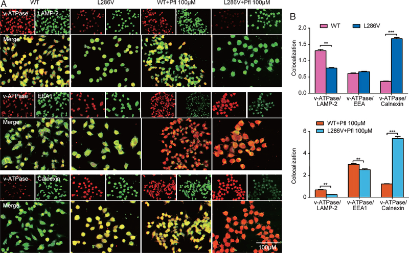
Fig. 3.
Propofol decreased lysosome vATPase in L286V Alzheimer’s disease cells by over activation of InsP3 (InsP3R) or ryanodine (RYR) receptors. A) Double-immunofluorescence labeling shows the colocalization of vATPase (V0a1 subunit) and lysosome (LAMP-2), or early endosome (EEA1), or ER (calnexin) in WT or L286V AD cells. Scale bar = 50 μm. B) Compared to WT controls, vATPase V0a1 subunit and lysosomal marker, LAMP-2 show little colocalization in L286V AD cells. Compared to WT controls, propofol at high concentration (Pfl 100 μΜ) significantly minimally colocalized vATPase VOal and lysosome marker, LAMP-2, but increased vATPase V0a1 in ER and early endosome. All data are expressed as the mean ± SEM from at least three separate experiments (N ≥ 3) of at least twenty individual cells and analyzed by unpaired t test followed by Tukey multiple comparison tests. *p < 0.01 and **p < 0.001.

