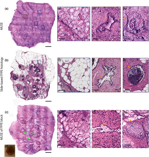
Fig. 8.
Breast tissue with human DCIS (green arrows) imaged with (a) MUSE, (b) slide-based FFPE histology, and (c) MUSE of an FFPE block face. A photo of the unstained tissue is as shown in (d). Zoomed-in features imaged with (e), (h), and (k) MUSE, (f), (i), and (l) slide-based FFPE histology, and (g), (j), and (m) MUSE of an FFPE block face. (e)–(g) Adipose tissue, (h)–(j) a benign duct, (k)–(m) DCIS. Scale bar: (a)–(c) 1 mm and (e)–(m) .

