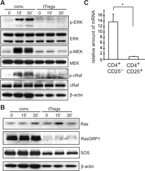
Figure 1.

Expression and activation of ERK signaling molecules in conventional T cells and tTregs. Ex vivo expanded CD4+CD25+ tTregs and conventional (conv) CD4+ T cells (CD4+ CD25- cells) isolated from C57BL/6 spleens were stimulated with biotin-conjugated anti-CD3 crosslinked with avidin for 0, 15, or 30 min. (A, B) tTregs and conv cells were analyzed by western blot analysis. Blots are representative of three independent experiments. (C) Real-time PCR assays for expression of Rasgrpl mRNAby conventional (CD4+CD25-) and tTregs (CD4+ CD25+). The relative amount of mRNA was determined using gapdh gene expression as a reference, *p<0.005 Student’s t-test. The data are representative of two independent experiments.
