Figure 2.

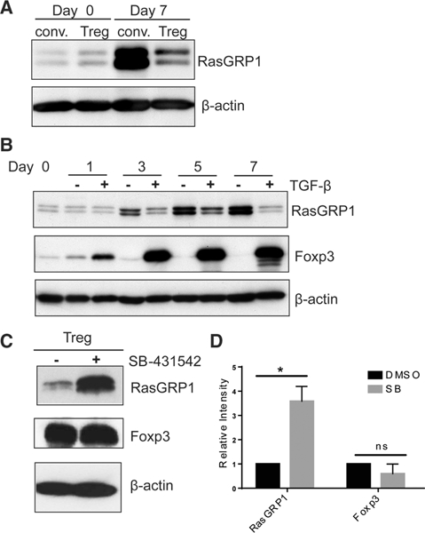
Regulation of RasGRPl expression by TGF-β. (A) CD4+ CD25+ Tregs and CD4+ CD25- conventional T cells were isolated from spleens of C57BL/6 mice and stimulated with anti-CD3/28 coated beads for 7 days in the presence of IL-2. Cells harvested at day 0 and day 7 were lysed in SDS sample buffer for western blot analysis using anti-RasGRPl and anti-p actin antibodies. (B) CD4+CD25- T cells were stimulated with plate-bound anti-CD3 and soluble anti-CD28 antibodies in the presence or absence of TGF-β supplemented with IL-2. After 3 days of stimulation, cells were harvested to remove stimulation and were further cultured in the presence of TGF-β and IL-2 and analyzed by western blot (C) CD4+25+ Tregs were expanded ex vivo for 7 days with anti-CD3 and anti-CD28 coated beads in the presence of IL-2. After 7 days, cells were re-stimulated on anti-CD3 and anti-CD28 coated plates in the presence of IL-2 and a TGF-β type I kinase inhibitor (SB-431542) or DMSO control. Cells were harvested 5 days after stimulation and lysed in SDS sample buffer then western blot analysis was performed using anti-RasGRPl, anti-Foxp3, and anti-p actin antibodies. (A-C) Blots are representative of three independent experiments. (D) Relative intensity of RasGRPl and Foxp3 in SB-43l542 treated Tregs compared to normalized DMSO control. β-actin was used as a loading control. Four spleens were pooled for each independent experiment. Data are shown as mean + SD (n = 3) and are representative of three independent experiments. Student’s t-test, RasGRPl p = 0.016; Foxp3 p = 0.157.
