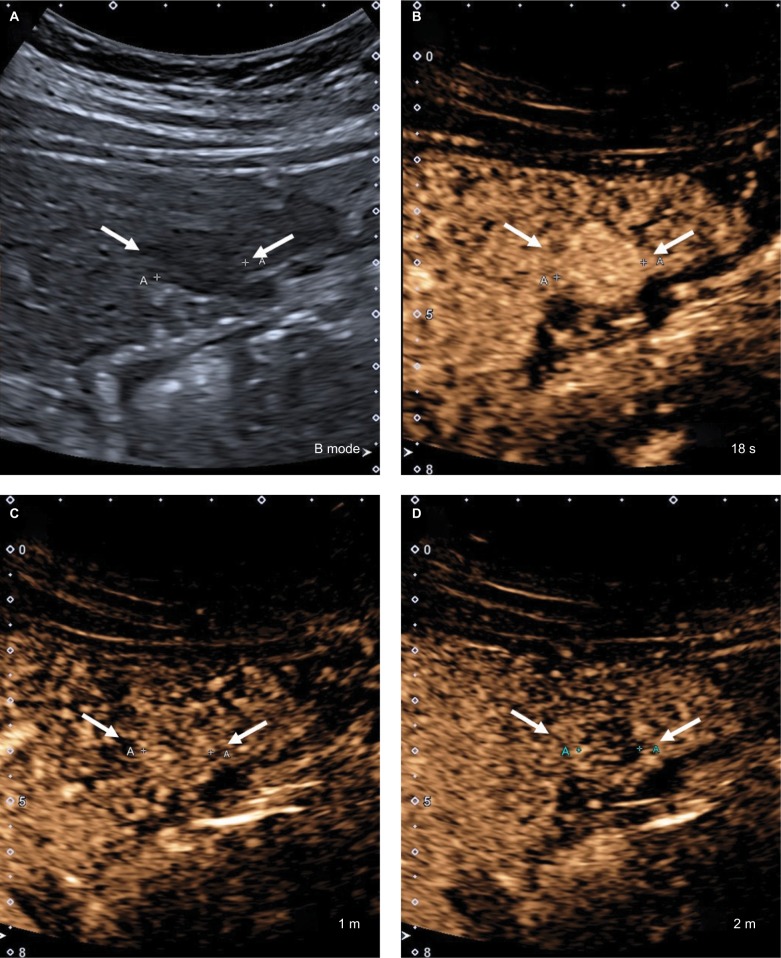
Figure 16.
CEUS LR-5.
Notes: (A) A 17 mm hypoechoic nodule on B-mode ultrasound. (B) The entire nodule shows hyperenhancement in the arterial phase. (C) At 1 minute, the nodule is isoenhancing compared with the surrounding liver parenchyma. (D) The nodule shows mild but definite hypoenhancement compared with the surrounding liver parenchyma at 2 minutes. This is late and mild washout. Arrows show the outline of the nodule.
Abbreviation: CEUS, contrast-enhanced ultrasound.

