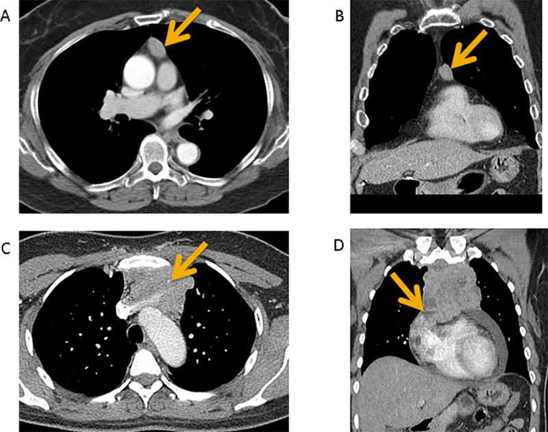
Figure 1 (A-D):

Contrast-enhanced CT of a pathologic IASLC/ITMIG TNM stage I (Masaoka-Koga stage IIA) type B2 thymoma demonstrates a prevascular mediastinal mass with smooth contours and homogeneous internal density in axial plane (A) and coronal plane (B). No invasive features are seen. Contrast-enhanced CT of a pathologic IASLC/ITMIG TNM stage IIIA (Masaoka-Koga stage III) type B3 thymoma demonstrates a prevascular mediastinal mass with lobulated contours and heterogeneous internal density in axial plane (C) and coronal plane (D). There is vascular endoluminal invasion of the innominate vein (C, arrow). There is abutment and also findings suggestive of invasion of adjacent mediastinal structures such as the pericardium (D, arrow).
