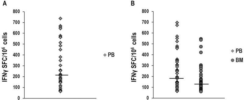
Figure 1. IFNγ-ELISPOT assay to investigate NPM1-mutated-specific T-cell responses.
Detection of NPM1-mutated-specific T cells producing IFNγ in peripheral blood (PB) and bone marrow (BM) samples, collected at different time-points from NPM1-mutated AML patients, after brief ex vivo stimulation (20 hours) with NPM1-mutated peptides. The ELISPOT assay, carried out after stimulation with a mixture containing all 18 NPM1-mutated (9–18 mers) peptides, documented NPM1-mutated-specific T cells in 34/52 (65.4%) PB samples (median 214 SFC/106 cells, range 63–736) (Panel A). NPM1-mutated-specific T cells were found by ELISPOT assay after stimulation with the combination of 13.9 and 14.9 peptides (Panel B), in 43/85 (50.6%) PB samples (median 194 SFC/106 cells, range 62–696) and in 34/80 (42.5%) BM samples (median 133 SFC/106 cells, range 62–546). Median absolute lymphocyte count observed in the analyzed BM samples was 1.9 × 10⁹/L (range 0.2–9.5). Black bars show median values. (P value > 0.05, Mann–Whitney U Test).

