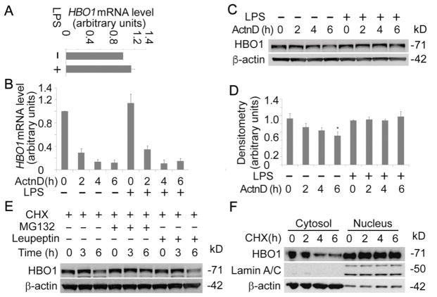
Fig. 2. LPS did not change HBO1 mRNA production and stability.
(A) Total RNA was isolated from untreated and LPS-treated THP-1 cells (125 ng/mL for 6 h). HBO1 mRNA levels were determined with qRT-PCR. (B) THP-1 cells were treated with Actinomycin D (5 μg/mL) or co-treated with Actinomycin D and LPS (125 ng/mL) for varying time points as indicated. Total RNA was extracted, and the HBO1 mRNA levels were determined using qRT-PCR. (C, D) Above treated cells were lysed, and cell lysates were subjected to HBO1 or β-actin immunoblotting. The densitometry results of C were plotted in D. (E) THP-1 cells were treated with CHX, CHX + MG132 or CHX + Leupeptin for varying time points as indicated. Cell lysates were subjected to HBO1 or β-actin immunoblotting. (F) THP-1 cells were treated with CHX for different time points, the cytoplasmic proteins and the nuclear proteins were separated and subjected to HBO1, Lamin A/C or β-actin immunoblotting. Lamin A/C was used as a nuclear marker, and β-actin was as a cytoplasmic marker. Data represents n=3 separate experiments. Graphs show mean ± SD and “*” denotes p<0.05.

