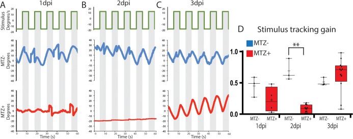
Fig 4. RPE ablation results in defects in visual function.
(A-C) To measure the OKR, the right eye of ablated and unablated larvae was exposed to a rotating stimulus, and the position of the stimulated eye was recorded at 1dpi (A, n = 3 unablated, n = 4 ablated), 2dpi (B, n = 3 unablated, n = 12 ablated) and 3dpi (C, n = 3 unablated, n = 12 ablated). (D) Quantification revealed that ablated larvae had a significantly reduced stimulus tracking gain at 2dpi (Mann-Whitney U test, P = 0.0055). Recovered OKR was detectible by 3dpi (Mann Whitney U test, P = 0.1878).

