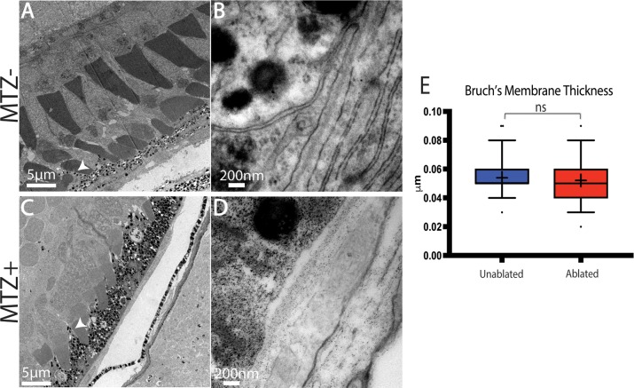
Fig 8. TEM analysis of regenerated RPE.
(A,B) TEM images of unablated 19dpf and (C,D) 14dpi eyes (C) Organized photoreceptor outer segments are visible in the ablated photoreceptor layer, and a regenerated RPE is present. (E) Quantification of BM thickness. Student’s T-test reveals that BM thickness is not significantly different in ablated larvae * p<0.05. (MTZ- n = 3 eyes, 81 measurements; MTZ+ n = 3, 81 measurements).

