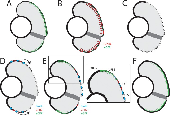
Fig 14. Model of RPE regeneration in larval zebrafish.
(A) nfsB-eGFP is specifically expressed in mature RPE in the central two-thirds of the eye. (B) Application of MTZ leads to apoptosis (red) of RPE and PRs. (C) RPE ablation leads to degeneration of PRs and Bruch’s Membrane (dotted line). (D) Unablated RPE in the periphery begin to proliferate and extend into the injury site (blue). (E) As regenerated eGFP+ RPE appear in the periphery, the RPE can be divided into 4 zones: peripheral RPE, differentiated RPE, transition zone, and injury site. (E, inset) Regenerated differentiated RPE appears in the periphery proximal to the unablated peripheral RPE, and contains proliferative cells adjacent to the transition zone. The transition zone consists of still-differentiating RPE cells and proliferative cells. The injury site is comprised of unpigmented proliferative cells that do not express any RPE differentiation markers. (F) Regeneration of a functional RPE layer and Bruch’s Membrane is complete by 14dpi.

