Figure 2.
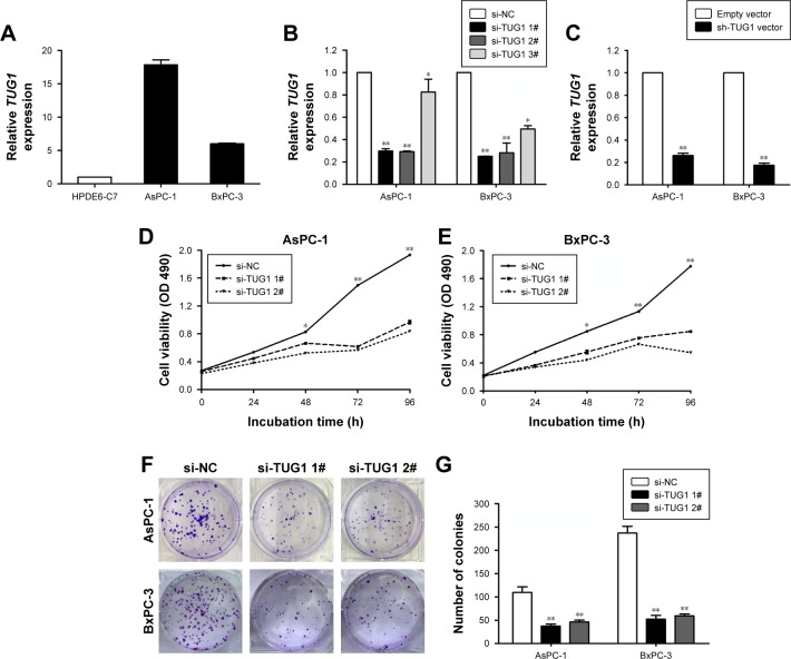
TUG1 promotes PC cell proliferation in vitro.
Notes: (A) Analysis of TUG1 expression levels in two PC cell lines (AsPC-1 and BxPC-3) compared with a normal pancreatic cell line (HPDE6-C7) using qRT-PCR. (B) The relative expression levels of TUG1 in AsPC-1 and BxPC-3 cells transfected with si-NC or si-TUG1 (si-TUG1 1#, si-TUG1 2#, and si-TUG1 3#) were measured using qPCR. (C) The relative expression levels of TUG1 in AsPC-1 and BxPC-3 cells transfected with an empty vector or sh-TUG1 were measured using qPCR. (D, E) MTT assays were performed to determine the cell viability for si-TUG1-transfected AsPC-1 and BxPC-3 cells. (F, G) Colony formation assays were used to determine the proliferation of si-TUG1-transfected AsPC-1 and BxPC-3 cells. *P<0.05, **P<0.01.
Abbreviations: PC, pancreatic cancer; TUG1, taurine upregulated 1; qRT, quantitative real-time; qPCR, quantitative PCR; NC, negative control.

