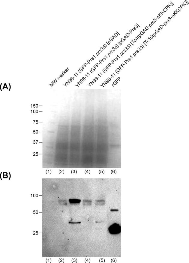
Figure 3.

Western blotting reveals that the deletion of 284KKCPK288 from Prs3 results in the loss of the GFP-signal associated with Prs1 in YN98–11. Crude extracts equivalent to 15 μg protein per lane were prepared from YN98–11 transformed with either pGAD (lane 2), pGAD-Prs3 (lane 3), Tc4(pGAD-prs3-ΔKKCPK) (lane 4) or Tc10(pGAD-prs3-ΔKKCPK) (lane 5) and separated on two simultaneously run 4–15% SDS-PAGE gels. (A) Coomassie Blue stained gel as loading control and (B) following transfer of the duplicated gel to a PVDF membrane western blotting was performed with specific anti-GFP antibodies (Santa Cruz sc-57587) and sc-2060 as primary and secondary antibodies, respectively. Successful electrophoresis and transfer to a PVDF membrane were checked by Ponceau S staining. The GFP signal was detected by chemiluminescence with anti-GFP antibodies. (A) Lane (1) molecular weight marker from Coomassie-stained gel. Lane (6) 10 μg rGFP standard.
